
200:1 Samples
May 19, 2017
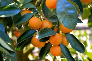
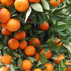
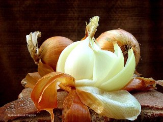
INTERSTELLAR PINE POLLEN
May 4, 2018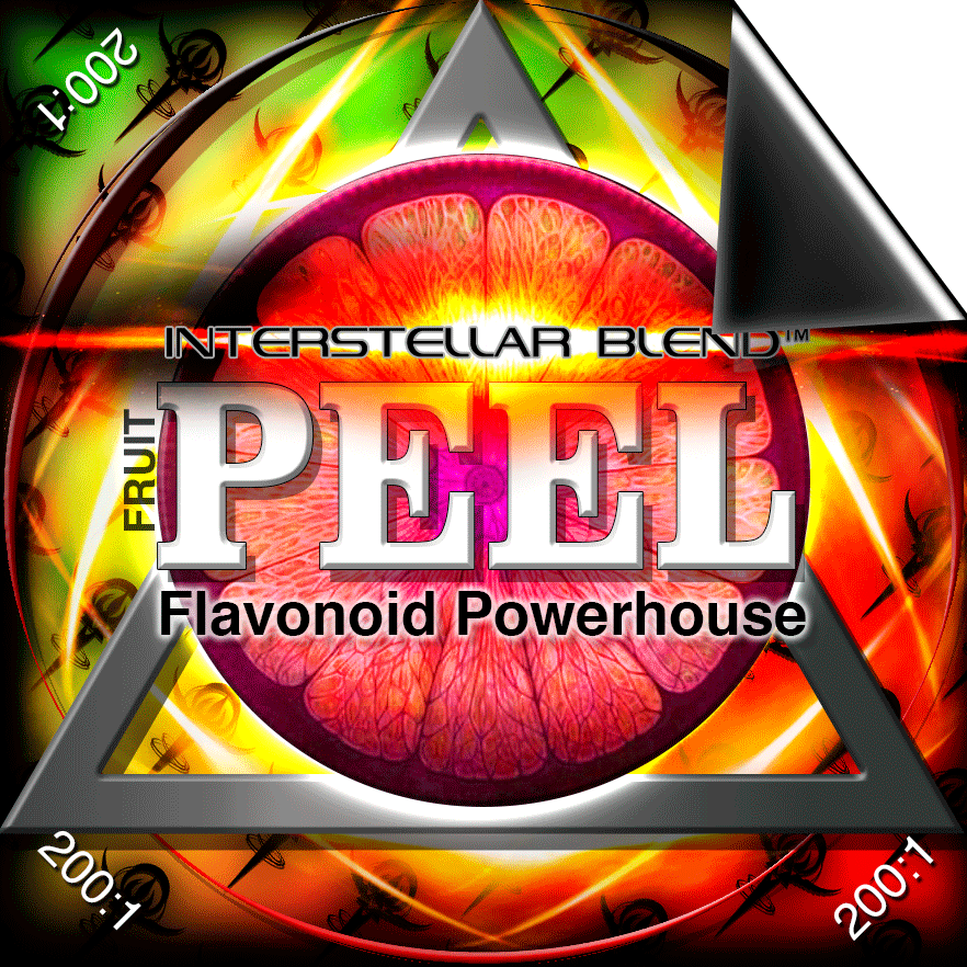
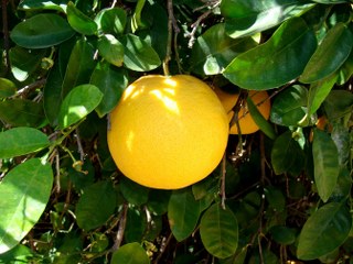
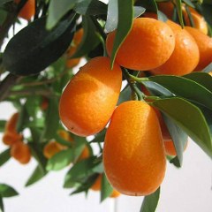
INTERSTELLAR PEEL BLEND
FLAVONOID POWERHOUSE
Insulin Sensitivity Restorer/ Diabetes Destroyer
200:1 100g = 300 1/8 tsp servings
20:1 100g = 36 1 tsp servings
*200:1 means it takes 200kg to make 1kg
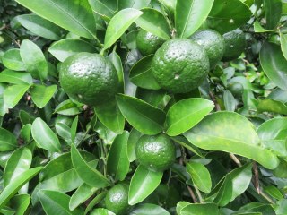
Fix the oxidative stress and you fix the insulin resistance. Diabetes is a byproduct of free radical damage. Flavonoids are the most powerful free radical scavengers on the planet. The vast majority of flavonoids are in the PEELS— the part everyone THROWS AWAY! No wonder diabetes and insulin resistance is rampant! Don’t believe me??? Check your blood sugar now and eat some orange peels for a few days and check again and you will see a HUGE improvement.
Increased oxidative stress precedes the onset of high-fat diet–induced insulin resistance and obesity
“We demonstrate that the pathways for reactive oxygen species (ROS) production and oxidative stress are coordinately up-regulated in both the liver and adipose tissue of mice fed an HFD before the onset of insulin resistance through discrete mechanism. “
FLAVONOIDS LOWER BLOOD PRESSURE
”Flavonoids from all 5 subgroups have been shown to attenuate a rise in or to reduce blood pressure during several pathological conditions (hypertension, metabolic syndrome, and diabetes mellitus). Flavones, flavonols, flavanones, and flavanols were able to modulate blood pressure by restoring endothelial function, either directly, by affecting nitric oxide levels, or indirectly, through other pathways.”
Oxidative stress shortens telomeres
“Telomeres in most human cells shorten with each round of DNA replication, because they lack the enzyme telomerase. This is not, however, the only determinant of the rate of loss of telomeric DNA. Oxidative damage is repaired less well in telomeric DNA than elsewhere in the chromosome, and oxidative stress accelerates telomere loss, whereas antioxidants decelerate it.”
Depression, anxiety-like behavior and memory impairment are associated with increased oxidative stress
“Co-occurrence of anxiety–depression like behaviors and memory deficits in rats correlates with elevated oxidative stress.”
Oxidative Stress and Anxiety: Relationship and Cellular Pathways
Anxiety is an alarm going off in your brain saying, “Yo!!! Yeah you!!! We got problems!!!”
“High O2 consumption, modest antioxidant defenses and a lipid-rich constitution make the brain highly vulnerable to redox imbalances. Oxidative damage in the brain causes nervous system impairment. Recently, oxidative stress has also been implicated in depression, anxiety disorders and high anxiety levels. The findings which establish a link between oxidative stress and pathological anxiety have inspired a number of other recent studies focusing on the link between oxidative status and normal anxiety and also on a possible causal relationship between cellular oxidative stress and emotional stress. This review examines the recent discoveries made on the link between oxidative status and normal anxiety levels and the putative role of oxidative stress in genesis of anxiety. We discuss the different opinions and questions that exist in the field and review the methodological approaches that are being used to determine a causal relationship between oxidative and emotional stress.”
Oxidative stress, cellular senescence and aging
“Almost a half century ago, the free radical theory of aging proposed that the reactive oxygen species (ROS) is a key component which contributes to the pathophysiology of aging in mammalian cells. Over the years, numerous studies have documented the role of oxidative stress caused by ROS in the aging process of higher organisms. In particular, several age-associated disease models suggest that ROS and oxidative stress modulate the incidence of age-related pathologies, and that it can strongly influence the aging process and possibly lifespan. The exact mechanism of ROS and oxidative stress-induced age-related pathologies is not yet very clear. Damage to biological macromolecules caused by ROS is thought to result in many age-related chronic diseases. At the cellular level, increased ROS leads to cellular senescence among other cellular fates including apoptosis, necrosis and autophagy. ”
Excessive caloric intake acutely causes oxidative stress
“These results suggest that the initial event caused by overnutrition may be oxidative stress, which produces insulin resistance, at least in part, via carbonylation and oxidation-induced inactivation of GLUT4.”
Oxidative stress and metabolic disorders
“Increased body weight and metabolic disorder including insulin resistance, type 2 diabetes and cardiovascular complications together constitute metabolic syndrome. The pathogenesis of metabolic syndrome involves multitude of factors. A number of studies however indicate, with some conformity, that oxidative stress along with chronic inflammatory condition pave the way for the development of metabolic diseases. Oxidative stress, a state of lost balance between the oxidative and anti-oxidative systems of the cells and tissues, results in the over production of oxidative free radicals and reactive oxygen species (ROS). Excessive ROS generated could attack the cellular proteins, lipids and nucleic acids leading to cellular dysfunction including loss of energy metabolism, altered cell signalling and cell cycle control, genetic mutations, altered cellular transport mechanisms and overall decreased biological activity, immune activation and inflammation. In addition, nutritional stress such as that caused by high fat high carbohydrate diet also promotes oxidative stress as evident by increased lipid peroxidation products, protein carbonylation, and decreased antioxidant system and reduced glutathione (GSH) levels. These changes lead to initiation of pathogenic milieu and development of several chronic diseases. Studies suggest that in obese person oxidative stress and chronic inflammation are the important underlying factors that lead to development of pathologies such as carcinogenesis, obesity, diabetes, and cardiovascular diseases through altered cellular and nuclear mechanisms, including impaired DNA damage repair and cell cycle regulation. Here we discuss the aspects of metabolic disorders-induced oxidative stress in major pathological conditions and strategies for their prevention and therapy.”
Acute oxidative stress and the ketogenic diet
“The mechanisms underlying the efficacy of the ketogenic diet (KD) remain unknown. Recently, we showed that the KD increased glutathione (GSH) biosynthesis. Since the NF E2-related factor 2 (Nrf2) transcription factor is a primary responder to cellular stress and can upregulate GSH biosynthesis, we asked whether the KD activates the Nrf2 pathway. Here we report that rats consuming a KD show acute production of H2O2 from hippocampal mitochondria, which decreases below control levels by 3 weeks, suggestive of an adaptive response. 4-Hydroxy-2-nonenal (4-HNE), an electrophilic lipid peroxidaytion end product known to activate the Nrf2 detoxification pathway, was also acutely increased by the KD. Nrf2 nuclear accumulation was evident in both the hippocampus and liver, and the Nrf2 target, NAD(P)H:quinone oxidoreductase (NQO1), exhibited increased activity in both the hippocampus and liver after 3 weeks. We also found chronic depletion of liver tissue GSH, while liver mitochondrial antioxidant capacity was preserved. These data suggest that the KD initially produces mild oxidative and electrophilic stress, which may systemically activate the Nrf2 pathway via redox signaling, leading to chronic cellular adaptation, induction of protective proteins, and improvement of the mitochondrial redox state.”
Autophagy as a Molecular Target of Flavonoids Underlying their Protective Effects in Human Disease.
“Autophagy is a cellular pathway with the ability to maintain cell homeostasis through the elimination of damaged or useless cellular components, and its deregulation may initiate or aggravate different human diseases. Flavonoids, a group of plant metabolites, are able to modulate different molecular and cellular processes including autophagy.
Analyzed publications indicated that imbalance between cell death and survival induced by changes in autophagy play an important role in the pathophysiology of a number of human diseases. The use of different flavonoids as autophagy modulators, alone or in combination with other molecules, might be a worthy strategy in the treatment of cancer, neurodegenerative disorders, cardiovascular diseases, hepatic diseases, leishmaniasis, influenza, gastric ulcers produced by Helicobacter pylori infection, diabetes, asthma, age-related macular degeneration or osteoporosis.”
RLIP76
When you turn off RLIP76 in mice they can’t get cancer, diabetes or become obese.
2-Hydroxyflavanone inhibits RLIP76 and can be found naturally in parsley, onion peels, berries, tea, and citrus fruit peels.
RLIP76 inhibition (via FLAVONOIDS IN orange peel) prevents Obesity, Metabolic syndrome and cancer
“Feeding a Western high-fat diet (HFD) to C57BL/6 mice induces obesity, associated with a chronic inflammatory state, lipid transport, and metabolic derangements, and organ system effects that particularly prominent in the kidneys. Here, we report that RLIP76 homozygous knock-out (RLIP76−/−) mice are highly resistant to obesity as well as these other features of metabolic syndrome caused by HFD. The normal increase in pro-inflammatory and fibrotic markers associated with HFD induced obesity in wild-type C57B mice was broadly and nearly completely abrogated in RLIP76−/− mice. This is a particularly striking finding because chemical markers of oxidative stress including lipid hydroperoxides and alkenals were significantly higher in RLIP76−/− mice. Whereas HFD caused marked suppression of AMPK in wild-type C57B mice, RLIP76−/−. The baseline renal function was reduced in mice had baseline activation of AMP-activated protein kinase, which was not further affected by HFDRLIP76−/− mice as compared with wild-type, but was unaffected by HFD, in marked contrast to severe renal impairment and glomerulopathy in the wild-type mice given HFD. Our findings confirm a fundamental role of RLIP76 in regulating the function of obesity-promoting pro-inflammatory cytokines, and provide a novel mechanism for targeted therapy of obesity and metabolic syndrome.”https://www.ncbi.nlm.nih.gov/pmc/articles/PMC3743508/
“When you get rid of this [RLIP76] gene in a mouse, it would appear that the mouse can’t get obese, it can’t get diabetes, it can’t get high cholesterol and it can’t get cancer,” explained Sanjay Awasthi, M.D., professor in the Division of Molecular Diabetes Research at City of Hope hospital.
RLIP76, a Glutathione-Conjugate Transporter, Plays a Major Role in the Pathogenesis of Metabolic Syndrome
Our recently published studies demonstrate that RLIP76−/− mice used for these studies were found to have marked insulin-sensitivity, and blood glucose was 46% lower than in RLIP76+/+animals (p<0.001). RLIP76−/− mice also had lower total serum cholesterol and triglycerides (43% and 40% of control, respectively; p<0.01) [1]. The hypoglycemia in RLIP76−/− mice is particularly remarkable because markers of oxidative-stress are remarkably increased in the tissues of the RLIP76−/− animals [1], [23]–[25]. Thus, in the absence of RLIP76, increases in these lipid-peroxidation products are insufficient by themselves to turn on any signaling pathway that can increase BG or lipids. Increased gluconeogenesis was particularly remarkable given that the activity of key gluconeogenic enzymes, G6Pase, F1,6-BPase, and PEPCK, in liver of RLIP76−/− mice was significantly inhibited.
Enhanced basal pAMPK levels in RLIP76−/− mice was another salient finding which strengthens the postulate that RLIP76 is a highly effective target for developing interventional strategies for MSy. Resveratrol, commonly used anti-oxidant, is known to activate AMPK which could contribute to its protective effects from high fat diet induced insulin-resistance [53], [54]. AMPK protects cells from stresses that cause ATP depletion by switching off ATP-consuming biosynthetic pathways. AMPK is activated by phosphorylation by an upstream protein kinase known as AMPK kinase. Activated AMPK can phosphorylate and regulate in vivo HMG-CoA, which is key regulatory enzyme of sterol synthesis [43], [47]. HMG-CoA limits the rate of cholesterol synthesis in liver tissue. Lipitor, inhibitor of HMG-CoA, exerts anti-inflammatory effects by lowering plasma cholesterol. Activation of AMPK leads to the inhibition of cholesterol synthesis by the phosphorylation of HMG-CoA reductase [43]. Loss of RLIP76 significantly affects the activation of stress and apoptosis pathway proteins [1], [25], [35]. Activation of AMPK leads to the inhibition of cholesterol synthesis by the phosphorylation of HMGCR. AMPK activation would be a good approach to treat T2D. These medications generally function to increase the effectiveness of insulin-mediated postprandial inhibition of hepatic gluconeogenesis. These findings provide a new insight on the mechanisms of action of hypoglycemic and/or hypolipidemic drugs.
RLIP76 knock-out mice survive well and are active. In our extensive and previously published studies, RLIP76 inhibition specifically leads to targeting signaling of importance in diabetes mellitus and other oxidative stress related conditions like cancers where targeting RLIP76 leads to selective cancer cell death without affecting the survival of normal cells and tissues [1],[31]–[33]. Hence, both global and selectively targeted approaches can be reasonably pursued as required while targeting RLIP76. In conclusion, our results suggest that RLIP76 is a key effector controlled by multiple proteins known to regulate the metabolic abnormalities of diabetes and metabolic syndrome, and that in its absence drugs that target these proteins will fail to function. The specific events that regulate the transport-effector/clathrin-endocytosis activity of RLIP76 (i.e. phosphorylation of RLIP76 by JNK, Akt, AMPK) will be explored in the future studies.
2′-Hydroxyflavanone (a flavonoid in orange peel): A novel strategy for targeting breast cancer
Intake of citrus fruits is known to reduce the risk for incidence of breast cancer. Hence, we tested the efficacy of citrus flavonoid 2′-hydroxyflavanone (2HF) in breast cancer. 2HF inhibited survival, clonogenic ability, cell cycle progression and induced apoptosis in breast cancer cells. 2HF also decreased VEGF levels and inhibited migratory capacity of breast cancer cells. Administration of 2HF led to regression of triple-negative MDA-MB-231 tumors in the mice xenograft model. 2HF decreased the levels of RLIP76 both studies and MDA-MB-231 xenograft model of breast cancer. Western blot and histopathological analyses of resected tumors showed a decline in the levels of survival and proliferation markers Ki67, pAkt, survivin, and cell cycle proteins CDK4 and cyclin B1. 2HF treatment led to inhibition of angiogenesis as determined by decreased VEGF levels and angiogenesis marker CD31 . 2HF reversed the pro-/anti-apoptotic ratio of BAX/BCL-2 by decreasing anti-apoptotic protein BCL-2 and increasing pro-apoptotic proteins BAX and BIM . 2HF also decreased the mesenchymal markers vimentin and fibronectin along with causing a parallel increase in pro-differentiation protein E-cadherin. Collectively, the ability of 2HF to decrease RLIP76, VEGF and regulate critical proliferative, apoptotic and differentiation proteins together provides strong rationale to further develop 2HF based interventions for targeting breast cancer.
It takes about 16 oranges to get a good dose of 2HF. Get the INTERSTELLAR PEEL BLEND instead in either 20:1 or 200:1 concentrations which is like eating that many oranges in just a few small scoops!
AWESOME MADE CONVENIENT!
INGREDIENTS:
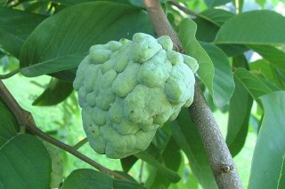
Annona cherimola Mill.(cherimoya) peel
Antihyperglycemic Activity of Annona cherimola Miller and Rutin on Alloxan-induced Diabetic Rats
“It can be concluded from the data that EEAc is beneficial in controlling the blood glucose level in experimental diabetic rats and according to in vivo studies it acts as α-glucosidase inhibitor. This effect can be attributed in part to the presence of flavonol glycoside, rutin. In addition, results validate the use of A. cherimola in Mexican traditional medicine for the treatment of diabetes. Finally, it is the first bioassay-guided about the chemistry and antidiabetic properties of A. cherimola.”
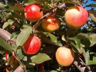 Apple peel extract (malus domestica)
“An increasing number of studies indicate that regular intake of fruits and vegetables have clear links to reduced risk of chronic diseases like diabetes and cardiovascular disease. The beneficial effects in many cases have been attributed to the phenolic and antioxidant content of the fruits and vegetables. Apples are a major source of fiber and contain good dietary phenolics with antioxidant function. Previous epidemiological studies have indicated that intake of apples reduces the risk of developing type 2 diabetes. Our studies indicate that this reduced risk is potentially because of the modulation of postprandial glucose increase by phenolics present in apples via inhibition ofα‐glucosidase. Phenolic content was evaluated during 3 months of postharvest storage of four varieties of apples and results indicated positive linkage to enhanced postharvest preservation andα‐glucosidase inhibition. These in vitro results along with existing epidemiological studies provide strong biochemical rationale for further animal or human clinical studies.”
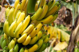 Banana peel (Musa Cavendish) extract
In vitro antioxidant, hypoglycemic and oral glucose tolerance test of banana peels
“Banana fruit is claimed to have antidiabetic effects despite its high calorie content, and its peels also contain vital phytoconstituents including gallocatechin. Previously banana pulp has been studied for antihyperglycemic effects, and in the present investigation antihyperglycemic effect of ethanolic extract of inner peels of Musa sapientum (EMS), Musa paradisiaca (EMP), Musa cavendish (EMC) and Musa acuminata (EMA) fruit was evaluated using oral glucose tolerance test in normoglycemic rats. In vitro antioxidant study was conducted using DPPH, H2O2 radical scavenging assay and ferric reducing power assay. Wistar rats were divided into fourteen groups and twelve groups received different doses of aforementioned extracts, while control group received gum acacia solution and remaining group received standard drug, glimepiride. All the rats received glucose load at a dose of 2 g/kg body weight. Groups treated with EMC and EMA showed significant decrease in glucose level (p < 0.01) at 150 min as compared to control group. In hypoglycemic study, only EMP 500 mg/kg, p.o. treated group revealed a significant decrease (p < 0.05) in glucose level at 120 min, while other groups did not show any sign of hypoglycemia. In glucose tolerance test, animals treated with EMC and EMA depicted dose dependent antihyperglycemic effect at 150 min while EMS and EMP showed significant reduction in plasma glucose at higher doses. In a similar fashion, EMA i.e. M. acuminata demonstrated highest antioxidant activity followed by EMC against DPPH radical. In ferric reducing power and H2O2 scavenging assay, EMA demonstrated maximal antioxidant activity when compared with other extracts.”
 Benincasa hispida peel extract
The Pharmacological Importance of Benincasa hispida
“Benincasa hispida phytochemical analysis showed that the major constituents of Benincasa hispida fruits are volatile oils , flavonoids, glycosides, sacchrides, proteins, carotenes, vitamins, minerals, ß-sitosterin and uronic acid. The pharmacological studies revealed that the plant exerted many pharmacological activities , including central nervous effects ( anxiolytic , muscle relaxant , antidepressant , in the treatment of Alzheimer’s disease and to minimize opiates withdrawal signs), antioxidant, anti-inflammatory, analgesic, antiasthmatic, diuretic , nephroprotective , antidiabetic , hypolipidemic and antimicrobial effects .”
 Bitter Orange peel extract (Citrus aurantium)
“Diabetes mellitus is a chronic disorder caused by partial or complete insulin deficiency, which produces inadequate glucose control and leads to acute and chronic complications. Premature and extensive arteriosclerosis involving renal, peripheral, and cardiovascular vessels remain the major complication of diabetes mellitus. Alteration in the serum lipid profile is known to occur in diabetes and this is likely to increase the risk for coronary heart disease. A reduction in serum lipids, particularly of the LDL and VLDL fraction and triglycerides, should be considered as being beneficial for the long-term prognosis of these patients.13 Lowering of blood glucose and plasma lipid levels through dietary modification and drug therapy seems to be associated with a decrease in the risk of vascular disease. In the present study, treatment with Citrus aurantium ethanolic extract (500 mg/kg b.w.) in normal rats produced significant decrease in blood glucose level. The hypoglycemic effect may be due to increased secretion of insulin from the b-cells of the pancreas, i.e., pancreatotrophic action.14 The results were comparable with that of tolbutamide, which acts by stimulation of insulin release,15 thus further confirming that the extract lowers the blood glucose by a similar action. Moreover, Citrus aurantium produced significant beneficial effects in the lipid profile in euglycemic rats, reducing triglycerides, total cholesterol, LDL, and VLDL, and increasing HDL, significantly. The ethanolic extract increased secretion of insulin from b-cells of pancreas; this increased secretion of insulin stimulates fatty acid biosynthesis and also the incorporation of fatty acids into triglycerides in the liver and adipose tissue.16 Alloxan, a beta cytotoxin, induces ‘chemical diabetes’ in a wide variety of animal species by damaging the insulin-secreting cells of the pancreas. Literature sources indicate that alloxan rats are hyperglycemic.17 The use of lower doses of alloxan (150 mg/kg b.w./i.p.) produced a partial destruction of pancreatic b-cells even though the animals became permanently diabetic. Thus, these animals have surviving b-cells and regeneration is possible.18 It is well known that the sulfonylureas (tolbutamide) act by directly stimulating the b-cells of the Islets of Langerhans More Details to release more insulin and these compounds are active in mild alloxan-induced diabetes where as they.17 Since our results show that tolbutamide reduced the blood glucose levels in the diabetic animals, the state of diabetes is not severe. Prolonged administration of an ethanolic extract of Citrus aurantium leads to significant reduction in blood glucose level, which is in agreement with other studies.18,20 The hypoglycemic activity of the drug was due to the regeneration of pancreatic cells that were partially destroyed by alloxan, and potentiation of insulin secretion from surviving b-cells of the islets of Langerhans.21 Diabetic rats were observed to have increased plasma lipids, which are responsible for several cardiovascular disorders.22 The higher lipid levels seen in diabetic rats was due to increased mobilization of free fatty acids from peripheral depots and also due to lipolysis caused by hormones.23,24 The ethanolic extract leads to regeneration of the b-cells of the pancreas and potentiation of insulin secretion from surviving b-cells; the increase in insulin secretion and the consequent decrease in blood glucose level may lead to inhibition of lipid peroxidation and control of lipolytic hormones. In this context, a number of other plants have also been reported to have antihyperglycemic, antihyperlipidemic, and insulin stimulatory effects.25,26,27 It is well known that LDL plays an important role in arteriosclerosis and that hypercholesterolemia is associated with a defect relating to the lack of LDL receptors. The decrease of cholesterol and LDL levels achieved by administration of ethanolic extract, demonstrates a possible protection against hypercholesterolemia and the harm this condition brings about. Further studies are needed to identify the chemical constituents of the ethanolic extract of Citrus aurantium that may be responsible for the hypoglycemic and hypolipidemic activity.”
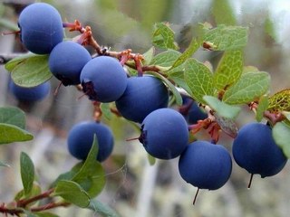 Blueberry peel extract
“This study was designed to evaluate cultivar variations in phenolic content, anthocyanin content, and antioxidant activity of peel and flesh; and to determine their potential inhibitory effects on α-glucosidase in 33 blueberry (Vaccinium species) cultivars, including 29 rabbiteye (Vaccinium ashei Reade) blueberries, two V. ashei hybrid derivatives, and two northern highbush (Vaccinium corymbosum L.). The relation of phenolic, anthocyanin, and antioxidant activity to α-glucosidase inhibition in blueberries also was investigated. It was found that peel tissue possessed higher levels of total anthocyanins (TA), total phenolics (TP), antioxidant capacity, and α-glucosidase inhibitory activity than flesh tissue in all blueberries tested. The percentage contributions of peel to whole berry on scavenging capacity against peroxyl free radicals (ROO
 Camellia sinensis fruit peel extract
“Tea fruit peel is the main byproduct during manufacture of tea seed oil. The great increase in tea seed oil production in recent years brings the challenge of finding application for tea fruit peel. The aims of this study were to obtain tea fruit peel extracts enriched with bioactive compounds by several solvent extraction methods and to evaluate their antioxidant activity and inhibitory potential against α-glucosidase and α-amylase in vitro. Flavonoids and phenolics were accumulated in ethyl acetate, butanol, and chloroform fractions, and these fractions possessed much better antioxidant activities, including scavenging effect on 1,1-diphenyl-2-picrylhydrazyl radicals (DPPH
► Tea fruit peel extracts could be used as natural antioxidant agents. ► Some fractions exhibited excellent inhibitory potential against α-glucosidase. ► Tea fruit peel can be developed as a renewable bioresource.”
Chinese Honey Orange peel extract (Citrus reticulata Blanco)
“Citrus reticulata Blanco cv. Murcot (honey) is one of the most important fruit tree crops in Pakistan. A detailed study regarding total antioxidant capacity (TAC), radical scavenging, antidiabetic and antimicrobial effects of the essential oils from leaves (CRL) and rind (CRR) of this plant were investigated. Essential oils obtained from steam distillation process were subjected to 2,2’-azinobis(3-ethylbenzothiazoline-6-sulpohonic acid) (ABTS) radical cation decolourization, ferric reducing antioxidant power (FRAP), 2.2’-Diphenyl-1-picrylhydrazil (DPPH) radical decolourization, lipid peroxide inhibition using linoleic acid emulsion, superoxide anion radical scavenging and iron chelation activity assays. A linear correlation was observed between the percent inhibition of ABTS radical cation and the amount of essential oils from leaves (R2 =0.9978) and rind (R2 = 0.985). The percent superoxide anion radical scavenging activity was found to be 15.25 and 48.75 for CRL and CRR, respectively. The reducing power in terms of FRAP values were found to be 1.2 and 0.173 mM FeSO4 for CRL and CRR, respectively. Both CRL and CRR exhibited good metal chelating activities, 56.10 and 61.82 for CRL and CRR, respectively. Best results regarding antidiabetic activity were shown by essential oils from leaves of Citrus reticulata with low as well as high concentrations. The oils also showed antibacterial and antifungal activities comparable to chloramphanicol. The data obtained from oils demonstrate the powerful antioxidative, radical scavenging, antidiabetic and antimicrobial properties of the plant. The data presented here shows that Citrus reticulata Blanco cv. Murcot (Honey) extracts have great antioxidant and radical scavenging activity and thus may be used as a good source of natural antioxidants. The in vivo efficacy of Citrus reticulata Blanco cv. Murcot (Honey) oils against diabetes mellitus or other degenerative diseases may be partially attributed to radical scavenging and antioxidant activity of the plant. “
 Citrus clementina peel extract
“This study reports the use of orange (Citrus × clementina) peel aqueous extract (OPE) for one-pot green synthesis of silver nanoparticles (AgNPs) with high effectiveness against various microbial pathogens as well as rat glial tumor C6 cells. The effects of various operational parameters on the synthesis of AgNPs were systematically investigated. The morphology, particle size, and properties of synthesized AgNPs were characterized using UV–visible spectroscopy, x-ray diffraction, x-ray photoelectron spectroscopy, field emission scanning electron microscopy, energy-dispersive x-ray spectroscopy, and Fourier transform infrared spectroscopy. High-resolution transmission electron microscopy shows that the nanoparticles are mostly spherical in shape and monodispersed, with an average particle size of 15–20 nm. Notably, the OPE-synthesized AgNPs were stable up to 6 months without change in their properties. Low doses of OPE-AgNPs inhibited the growth of human pathogens Escherichia coli, Bacillus cereus, andStaphylococcus aureus. The minimum inhibitory concentration and minimum bactericidal concentration of AgNPs against selected pathogenic bacteria were determined. OPE-AgNPs exhibited strong antioxidant activity in terms of ABTS (2,2′-azino-bis (3-ethylbenzothiazoline-6-sulfonic acid)) radical scavenging (IC50 49.6 μg/mL) and DPPH (1,1-diphenyl-2-picrylhydrazyl) radical scavenging (IC5063.4 μg/mL). OPE-AgNPs showed dose-dependent response against rat glial tumor C6 cells (LD50 60 μg/mL) showing a promising potential as anticancer agents.”
Citrus depressa Hayata (shiikuwasa) peel extract
“Citrus depressa Hayata (commonly known as shiikuwasa) is cultivated in the northern areas of Okinawa, Japan, and used as a juice. In this study, we examined the anti-obesity effects and mechanism of action of shiikuwasa peel extract (SE) using high-fat diet (HFD)-induced obese mice. Mice were fed a low-fat diet (LFD), HFD or HFD containing 1% or 1.5% (w/w) SE (HFD + 1 SE and HFD + 1.5 SE, respectively) for 5 weeks. The body weight gain and white adipose tissue weight were significantly decreased in the HFD + 1.5 SE group compared with the HFD group. The plasma triglyceride and leptin levels were also significantly reduced in the HFD + 1.5 SE group compared with the HFD group. Histological examinations showed that the sizes of the adipocytes were significantly smaller in the HFD + 1.5 SE group than in the HFD group. The HFD + 1.5 SE group also showed significantly lower mRNA levels of lipogenesis-related genes, such as activating protein 2, stearoyl-CoA desaturase 1, acetyl-CoA-carboxylase 1, fatty acid transport protein and diacylglycerol acyltransferase 1, than the HFD group. These results suggest that the anti-obesity effects of SE may be elicited by regulating the expressions of lipogenesis-related genes in white adipose tissue.”
 Citrus medica L. cv Diamante peel extract (rutaceae)
“This study aimed to investigate the antidiabetic, antilipidaemic and antioxidant activities of Citrus medica cv Diamante (Rutaceae) hydroalcoholic (CD) peel extract in Zucker diabetic fatty (ZDF) rats. The ability of CD to protect against oxidative stress was investigated by using different in vitro assays and in vivo by using the reactive oxygen metabolites-derived compounds (d-ROMs) test and the biological antioxidant potential test (BAP). Two different doses of CD extract (300 and 600 mg/kg/die) were administered at ZDF rats for 4 weeks. CD reduced cholesterol and triglycerides levels. A dose-dependent effect on body weight and serum glucose levels was observed. A decrease of d-ROMs and an increase of BAP were recorded by using the dose of 600 mg/kg. The extract inhibited lipid peroxidation (IC50 value of 0.23 mg/ml). These findings suggest as an efficient phytotherapeutic approach in combating hyperlipidaemic and hyperglycaemic disorders.”
Cucumis sativus peel EXCTRACT
Protective Role of Three Vegetable Peels in Alloxan Induced Diabetes Mellitus in Male Mice
“In a preliminary study, effects of ethanolic extracts of Cucurbita pepo, Cucumis sativus andPraecitrullus fistulosus peels were studied at 250 and 500 mg kg−1 d−1 for 15 days in the alterations in serum glucose and in hepatic lipid peroxidation (LPO) in male mice. In the pilot experiment, the effective and safe concentration of each peel was administered (p.o.) for 10 consecutive days and then on 11th and 12th days alloxan was administered along with peel extracts. The treatment was continued up to 15th day. At the end, alterations in serum glucose, insulin, triiodothyronine, thyroxine, total cholesterol, triglyceride, high density lipoprotein, low density lipoprotein, very low density lipoprotein, hepatic lipid peroxidation, superoxide dismutase and catalase were studied. All the three peel extracts nearly reversed most of these changes induced by alloxan suggesting their possible role in ameliorating diabetes mellitus and related changes in serum lipids. Total polyphenols, flavonoids and ascorbic acid contents of the test peels were also estimated, which appear to be associated with the observed antidiabetic and antioxidative potentials.”
 Cucurbita pepo FRUIT peel extract
“This study was aimed to evaluate the hypoglycaemic and hypolipidemic effects of different doses of pumpkin (Cucurbita pepo L.) powder in male diabetic rats. A total of 35 rats were randomized into 5 groups of 7 each as follows: Group 1: Normal control; Group 2: Diabetic control; Group 3: Diabetics administered with low doses of pumpkin powder (1 g/kg); Group 4: Diabetics administered with high doses of pumpkin powder (2 g/kg), and Group 5: Diabetics administered with glibenclamide (0.6 mg/kg), as positive control. The rats were made diabetic by alloxan (120 mg/kg body weight (BW)) injection and were treated for 4 weeks on a daily basis. Blood samples were collected following the experiment. Pancreatic specimens were also collected for histological analysis. Glucose, cholesterol, triglycerides, low density lipoprotein (LDL) and C-reactive protein (CRP) were significantly increased, while insulin was decreased in diabetic rats as compared to the normal control group (p < 0.05). Low dose pumpkin significantly decreased glucose, triglycerides, LDL and CRP as compared to diabetic group and high dose pumpkin decreased cholesterol (p < 0.05). Histological analysis also revealed a significant increase in the diameter and number of langerhans islets in treated group with pumpkin, which was consistent with the latter findings. Therefore, pumpkin might be beneficial in diabetic patients.”
 Dracaena cochinchinensis resin (dragons blood)
“Dragon’s blood” is the name given to a deep red resin obtained from a variety of plant sources. The resin extracted from stems of Dracaena cochinchinensis is one such source of “dragon’s blood”. It has a reputation for facilitating blood circulation and dispersing blood stasis. In traditional Chinese medicine, this resinous medicine is commonly prescribed to invigorate blood circulation for the treatment of traumatic injuries, blood stasis and pain. Modern pharmacological studies have found that this resinous medicine has anti-bacterial, anti-spasmodic, anti-inflammatory, analgesic, anti-diabetic, and anti-tumor activities, while it is also known to enhance immune function, promote skin repair, stop bleeding and enhance blood circulation. Various compounds have been isolated from the plant, including loureirin A, loureirin B, loureirin C, cochinchinenin, socotrin-4′-ol, 4′,7-dihydroxyflavan, 4-methylcholest-7-ene-3-ol, ethylparaben, resveratrol, and hydroxyphenol. The present review summarizes current knowledge concerning the botany, phytochemistry, pharmacological effects, toxicology studies and clinical applications of this resinous medicine as derived from D. cochinchinenesis.”
 Durian (Durio zibethinus Murr.) peel extract
“Durian contains flavonoids, namely catechin and quercetin [23] as well as polyphenols and tannins [10]. Quercetin had activity Aldos reductase inhibitors that could potentially be used in therapeutic antihyperglycemia [24]. In the rambutan fruit peels contained flavonoids and tannins [12]. Ethanolic extract of rambutan fruit peels contained quercetin, geraniin [13] and epigallocatechin-3-gallate (EGCG) which had antihyperglycemia activity [14] as well as powerful antioxidants [15]. Based on the content of flavonoids catechin, quercetin and EGCG and polyphenols and tannins, a mechanism thought to decrease blood glucose levels in diabetic rats through inhibition of glucose absorption, stimulates the release of insulin and indirect mechanisms through the antioxidant processes. Glucose binds to proteins can be oxidized and produce Reactive Oxygen Species (ROS). The combination of glycation and oxidation of glucose produces AGEs (advanced glycogen end-products) in which this process was irreversible long-lasting and can caused tissue damage. That glycated proteins and AGEs-modified proteins can lead to oxidative stress which can trigger the diabetic condition [22]. Another mechanism that was thought to have the effect of decreasing blood glucose levels in test animals, with the occurrence geraniin in rambutan fruit peel extract which had the ability to prevent the formation of AGEs [13]. This research has proven that the ethanol extract of durian rind and rambutan fruit peel can lowered blood glucose levels of alloxan-induced rats. However, the mechanism of the antidiabetic activity of the ethanol extract of durian and rambutan fruit peels was not certainly. So the evaluation to determine the mechanism of molecular pharmacology decrease in blood glucose levels that occurs, needs to be done. In addition it is necessary to ensure what the active compounds are most responsible for its pharmacological activity.”
 Grapefruit peel extract (Citrus paradisi)
“This study sought to characterize the interaction of phenolic extract from grapefruit peels with α‐amylase, α‐glucosidase and angiotensin‐I‐converting enzyme (ACE) activities. The free phenolic of grapefruit peels was extracted with 80% acetone, while the bound phenolic was extracted from the alkaline and acid‐hydrolyzed residue with ethyl acetate; and their interaction with the enzymes were assessed. The phenolic extracts inhibited α‐amylase, α‐glucosidase and ACE enzyme activities; however, free phenolic had significantly higher (P < 0.05) α‐amylase and α‐glucosidase inhibitory activities than bound phenolic, while there was no significant difference (P > 0.05) in their ACE inhibitory activities. Nevertheless, the extracts were strong inhibitor of α‐glucosidase when compared to α‐amylase. The phenolic inhibited sodium nitroprusside‐induced lipid peroxidation in pancreas in a dose‐dependent manner. Therefore, free and bound phenolic extracts from grapefruit peels could be used as nutraceutical for the management of hypertension and type 2 diabetes.
Type 2 diabetes (including its precursor, metabolic syndrome) is one of the top five most significant diseases in the developed world and is rapidly becoming a burden in sub‐Sahara Africa. Natural inhibitors with relatively mild inhibition of α‐amylase but strong inhibitor of α‐glucosidase activity could be an effective therapy for type 2 diabetes with minimal side effects, while the inhibition of angiotenisn‐I‐converting enzyme is a useful approach in the treatment of hypertension in both diabetic and nondiabetic patients, and phenolics have promising potential. Our findings in this study have shown that grapefruit peels that are usually considered to be waste could be a cheap and good source of free soluble and bound phenolics with antidiabetic and antihypertensive potentials.”
Hawthorn (Crataegus Mexicana) peel extract
“This study provides the first in vitro evidence for an active role of C. mexicana as antioxidant on erythrocytes. Hawthorn is a fruit with anti-oxidant potential, reduces morphological changes and extends life span of the red blood cells in vitro. Such protection could be due to the inhibition of membrane lipoperoxidation and oxidative damage in cytoskeletal proteins. The conjunction of antioxidants in this extract, and not just specific purified antioxidants, could be helpful to keep normal the rheology properties of the erythrocyte apparently without any pro-oxidative damage.
The antioxidants present in the acetone extract may contribute to protect the erythrocyte cell membrane against lipid peroxidation induced by oxidative stress, thereby participating in diminishing the morphological and many rheology changes that may happen in the pathogenesis of vasoocclusive diseases, such diabetes mellitus, metabolic syndrome, different types of anemia, and the inhibition of cell storage lesion in RBCs.
More studies are required to correctly identify the bioactive compounds of C. mexicana and how it contribute to the erythrocyte protection, and their mechanisms of action in the protection of the RBCs. This study give the scientific biochemical bases to use hawthorn extracts in a clinical setting, to prevent or treat many diseases and conditions associated to oxidative stress.”
 Huyou (Citrus changshanensis) fruit peel extract
“Huyou (Citrus changshanensis) is rich in naringin and neohesperidin, which are natural flavanone glycosides with a range of biological activities. Among the different fruit parts, i.e. flavedo, albedo, segment membrane (SM), and juice sacs (JS), albedo showed the highest contents of both compounds, with 27.00 and 19.09 mg/g DW for naringin and neohesperidin, respectively. Efficient simultaneous purification of naringin and neohesperidin from Huyou albedo was established by the combination of macroporous D101 resin chromatography and high-speed counter-current chromatography (HSCCC). Purified naringin and neohesperidin were identified by both HPLC and LC–MS, and their effects on glucose consumption were investigated in HepG2 cells. Cells treated with naringin and neohesperidin showed increased consumption of glucose, and this was associated with increased phosphorylation of AMP-activated protein kinase (AMPK). Therefore, naringin and neohesperidin from Huyou may act as potential hypoglycaemic agents through regulation of glucose metabolism.”
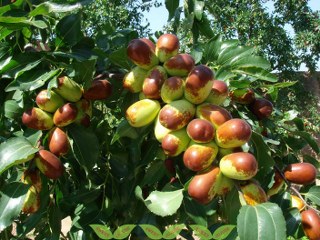 Jujube (Ziziphus Jujuba Mill.) Fruit peel
“The nutritional jujube (Ziziphus jujube Mill.) fruit belonging to the Rhamnaceous family grows mostly in Europe, southern and eastern Asia, and Australia, especially the inland region of northern China. Jujube has a long history of usage as a fruit and remedy. The main biologically active components are vitamin C, phenolics, flavonoids, triterpenic acids, and polysaccharides. Recent phytochemical studies of jujube fruits have shed some light on their biological effects, such as the anticancer, anti-inflammatory, antiobesity, immunostimulating, antioxidant, hepatoprotective, and gastrointestinal protective activities and inhibition of foam cell formation in macrophages. A stronger focus on clinical studies and phytochemical definition of jujube fruits will be essential for future research efforts. This review may be useful for predicting other medicinal uses and potential drug or food interactions and may be beneficial for people living where the jujube fruits are prevalent and health care resources are scarce.”
 Kumquat (Fortunella margarita ) peel
“Obesity is a nutritional disorder associated with many health problems such as dyslipidemia, type 2 diabetes and cardiovascular diseases. In the present study, we investigated the anti-metabolic disorder effects of kumquat (Fortunella margarita Swingle) fruit extract (FME) on high-fat diet-induced C57BL/6 obese mice.
The kumquat fruit was extracted with ethanol and the main flavonoids of this extract were analyzed by HPLC. For the preventive experiment, female C57BL/6 mice were fed with a normal diet (Chow), high-fat diet (HF), and high-fat diet with 1% (w/w) extract of kumquat (HF+FME) for 8 weeks. For the therapeutic experiment, female C57BL/6 mice were fed with high-fat diet for 3 months to induce obesity. Then the obese mice were divided into two groups randomly, and fed with HF or HF+FME for another 2 weeks. Body weight and daily food intake amounts were recorded. Fasting blood glucose, glucose tolerance test, insulin tolerance test, serum and liver lipid levels were assayed and the white adipose tissues were imaged. The gene expression in mice liver and brown adipose tissues were analyzed with a quantitative PCR assay.
In the preventive treatment, FME controlled the body weight gain and the size of white adipocytes, lowered the fasting blood glucose, serum total cholesterol (TC), serum low density lipoprotein cholesterol (LDL-c) levels as well as liver lipid contents in high-fat diet-fed C57BL/6 mice. In the therapeutic treatment, FME decreased the serum triglyceride (TG), serum TC, serum LDL-c, fasting blood glucose levels and liver lipid contents, improved glucose tolerance and insulin tolerance. Compared with the HF group, FME significantly increased the mRNA expression of PPARα and its target genes.
Our study suggests that FME may be a potential dietary supplement for preventing and ameliorating the obesity and obesity-related metabolic disturbances.”
 Lagenaria siceraria peel extract
” In conclusion, our present findings reveal that the peel extract of Lagenaria siceraria, not only regulates lipid peroxidation, but also appears to be a promising agent for the amelioration of hyperthyroidism or hyperglycemia.”
 Lansium domesticum fruit peel extract
“The peel of L. domesticum fruits possessed higher O2−• and OH• scavenging activity than the seeds, particularly when extracted with 50% aqueous ethanol and partitioned with ethyl acetate (LDSK50-EA). This fraction had high potential of both hydrophilic and lipophilic antioxidants. Moreover, LDSK50-EA had a geno-protective effect by reduction of the DNA damage induced by H2O2 radicals, proven by comet assay in TK6 cells. This study generated new and updated information on the biological activity of extracts of long-kong fruits. It may lead to a discovery of new alternative sources of natural antioxidant and anti-genotoxic substances for the prophylaxis or treatment of free radical-related diseases as well as the development of the nutraceutical product industry.”
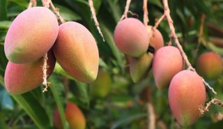 Mango peel extract (Mangifera indica)
“In the present study, the composition of mango peel powder (MPP) collected from the mango pulp industry was determined and the effect of MPP on ameliorating diabetes and its associated complications was studied.
Mango peel was rich in polyphenols, carotenoids and dietary fibre. Peel extract contained various bioactive compounds and was found to be rich in soluble dietary fibre. Peel extract exhibited antioxidant properties and protected against DNA damage. Therefore, the effect of peel on ameliorating diabetes was investigated in a rat model of diabetes. A significant increase in urine sugar, urine volume, fasting blood glucose, total cholesterol, triglycerides and low density lipoprotein, and decrease in high density lipoprotein were observed in the rats; however, these parameters were ameliorated in diabetic rats fed with diet supplemented with mango peel at 5% and 10% levels in basal diet. Treatment of diabetic rats with MPP increased antioxidant enzyme activities and decreased lipid peroxidation in plasma, kidney and liver compared to untreated diabetic rats. Glomerular filtration rate and microalbuminuria levels were ameliorated in MPP treated diabetic group.
Mango peel, a by‐product, can be used as an ingredient in functional and therapeutic foods.”
 Meyer Lemon peel extract (Citrus × meyeri)
Protective effects of lemon flavonoids on oxidative stress in diabetic rats
“The effects of lemon flavonoids, as crude flavonoids prepared from lemon juice, were investigated in diabetic rats. The oxidative stress of eriocitrin (eriodictyol 7‐O‐β‐rutinoside) and hesperidin (hesperetin 7‐O‐β‐rutinoside) on streptozotocin‐induced diabetic rats was investigated. Diabetic rats were given a diet which contained 0.2% crude flavonoids, 0.2% eriocitrin, and 0.2% hesperidin. After the 28‐d feeding period, the concentration of the thiobarbituric acid‐ reactive substance in the serum, liver, and kidney of diabetic rats administered crude flavonoids, eriocitrin, and hesperidin significantly decreased as compared with that of the diabetic group. The levels of 8‐hydroxydeoxyguanosine, which is exchanged from deoxyguanosine owing to oxidative stress, in the urine of diabetic rats administered eriocitrin and hesperidin significantly decreased as compared with that of the diabetic rat group. Crude flavonoids, eriocitrin, and hesperidin suppressed the oxidative stress in the diabetic rats. These results demonstrated that dietary lemon flavonoids of eriocitrin and hesperidin play a role as antioxidant in vivo.”
 Momordica charantia fruit peel extract
“We studied the long‐term effects of streptozotocin‐induced diabetes on tissue‐specific cytochrome P450 (CYP) and glutathione‐dependent (GSH‐dependent) xenobiotic metabolism in rats. In addition, we also studied the effect of antidiabetic Momordica charantia (karela) fruit‐extract feeding on the modulation of xenobiotic metabolism and oxidative stress in rats with diabetes. Our results have indicated an increase (35–50%) in CYP4A‐dependent lauric acid hydroxylation in liver, kidney, and brain of diabetic rats. About a two‐fold increase in CYP2E‐dependent hepatic aniline hydroxylation and a 90–100% increase in CYP1A‐dependent ethoxycoumarin‐O‐deethylase activities in kidney and brain were also observed. A significant increase (80%) in aminopyrene N‐demethylase activity was observed only in rat kidney, and a decrease was observed in the liver and brain of diabetic rats. A significant increase (77%) in NADPH‐dependent lipid peroxidation (LPO) in kidney of diabetic rats was also observed. On the other hand, a decrease in hepatic LPO was seen during chronic diabetes. During diabetes an increased expression of CYP1A1, CYP2E1, and CYP4A1 isoenzymes was also seen by Western blot analysis. Karela‐juice feeding modulates the enzyme expression and catalytic activities in a tissue‐ and isoenzyme‐specific manner. A marked decrease (65%) in hepatic GSH content and glutathione S‐transferase (GST) activity and an increase (about two‐fold) in brain GSH and GST activity was observed in diabetic rats. On the other hand, renal GST was markedly reduced, and GSH content was moderately higher than that of control rats. Western blot analyses using specific antibodies have confirmed the tissue‐specific alterations in the expression of GST isoenzymes. Karela‐juice feeding, in general, reversed the effect of chronic diabetes on the modulation of both P450‐dependent monooxygenase activities and GSH‐dependent oxidative stress related LPO and GST activities. These results have suggested that the modulation of xenobiotic metabolism and oxidative stress in various tissues may be related to altered metabolism of endogenous substrates and hormonal status during diabetes. The findings may have significant implications in elucidating the therapeutic use of antidiabetic drugs and management of Type 1 diabetes in chronic diabetic patients.”
 Niihime (Citrus unshiu Citrus tachibana) fruit peel
“Niihime is a sour Citrus fruit produced along the coast of the Sea of Kumano in the Mie prefecture of Japan. A novel ferulate derivative was isolated from niihime fruit. The structure of the compound was elucidated by spectroscopic analyses and identified as saccharic acid 1,4-lactone 3,5-di-O-ferulate by 1H-NMR, 13C-NMR, and MS analyses. The new compound exhibited significantly higher antioxidative activity than ferulic acid, a parent molecule of the compound, and trolox, a standard antioxidant, as measured by an ORAC assay (P < 0.05). Further, it was abundantly contained in the flavedo, albedo, and segment epidermis of the niihime peel.”
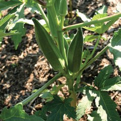 Okra (Abelmoschus esculentus (L.) Moench) peel
The present investigation was aimed to study the antidiabetic and antihyperlipidemic potential ofAbelmoschus esculentus peel and seed powder (AEPP and AESP) in streptozotocin (STZ)-induced diabetic rats.
Acute toxicity of AEPP and AESP was studied in rats at 2000 mg/kg dose and diabetes was induced in rats by administration of STZ (60 mg/kg, i.p.). After 14 days of blood glucose stabilization, diabetic rats received AEPP, AESP, and glibenclamide up to 28 days. The blood samples were collected on day 28 to estimate the hemoglobin (Hb), glycosylated hemoglobin (HbA1c), serum glutamate-pyruvate transferase (SGPT), total protein (TP), and lipid profile levels.
In acute toxicity study, AESP and AESP did not show any toxicity or death up to a dose of 2000 mg/kg. Therefore, to assess the antidiabetic action, one by fifth and one by tenth dose of both powders were selected. Administration of AEPP and AESP at 100 and 200 mg/kg dose in diabetic rats showed significant (P < 0.001) reduction in blood glucose level and increase in body weight than diabetic control rats. A significant (P < 0.001) increased level of Hb, TP, and decreased level of HbA1c, SGPT were observed after the treatment of both doses of AEPP and AESP. Also, elevated lipid profile levels returned to near normal in diabetic rats after the administration of AEPP and AESP, 100 and 200 mg/kg dose, compared to diabetic control rats.
The present study results, first time, support the antidiabetic and antihyperlipidemic potential of A. esculentus peel and seed powder in diabetic rats.
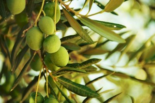 Olive leaf (Olea europaea)
“This study was designed to test the antidiabetic and antioxidative activities of olive leaf oleuropein and hydroxytyrosol. Diabetes in Wistar rats was induced by intraperitoneal injections of alloxan. The serum glucose and cholesterol, hepatic glycogen, the thiobarbituric acid-reactive substances (TBARS), and the components of hepatic and serum antioxidant system were examined. Diabetic rats showed hyperglycemia, hypercholesterolemia, increased lipid peroxidation, and depletion in the antioxidant enzymes activities. The administration, for 4 weeks, of oleuropein and hydroxytyrosol rich extracts, leading to 8 and 16 mg/kg body weight of each compound, significantly decreased the serum glucose and cholesterols levels and restored the antioxidant perturbations. These results suggested that the antidiabetic effect of oleuropein and hydroxytyrosol might be due to their antioxidant activities restraining the oxidative stress which is widely associated with diabetes pathologies and complications.”
 Onion peel extract (Allium cepa)
“Quercetin derivatives in onions have been regarded as the most important flavonoids to improve diabetic status in cells and animal models. The present study was aimed to examine the hypoglycemic and insulin-sensitizing capacity of onion peel extract (OPE) containing high quercetin in high fat diet/streptozotocin-induced diabetic rats and to elucidate the mechanism of its insulin-sensitizing effect.
Male Sprague-Dawley rats were fed the AIN-93G diet modified to contain 41.2% fat and intraperitoneally injected with a single dose of streptozotocin (40 mg/kg body weight). One week after injection, the rats with fasting blood glucose levels above 126 mg/dL were randomly divided into 4 groups to treat with high fat diet containing 0 (diabetic control), 0.5, or 1% of OPE or 0.1% quercetin (quercetin equivalent to 1% of OPE) for 8 weeks. To investigate the mechanism for the effects of OPE, we examined biochemical parameters (insulin sensitivity and oxidative stresses) and protein and gene expressions (pro-inflammatory cytokines and receptors).
Compared to the diabetic control, hypoglycemic and insulin-sensitizing capability of 1% OPE were demonstrated by significant improvement of glucose tolerance as expressed in incremental area under the curve (P = 0.0148). The insulin-sensitizing effect of OPE was further supported by increased glycogen levels in liver and skeletal muscle (P < 0.0001 and P = 0.0089, respectively). Quantitative RT-PCR analysis showed increased expression of insulin receptor (P = 0.0408) and GLUT4 (P = 0.0346) in muscle tissues. The oxidative stress, as assessed by superoxide dismutase activity and malondialdehyde formation, plasma free fatty acids, and hepatic protein expressions of IL-6 were significantly reduced by 1% OPE administration (P = 0.0393, 0.0237, 0.0148 and 0.0025, respectively).
OPE might improve glucose response and insulin resistance associated with type 2 diabetes by alleviating metabolic dysregulation of free fatty acids, suppressing oxidative stress, up-regulating glucose uptake at peripheral tissues, and/or down-regulating inflammatory gene expression in liver. Moreover, in most cases, OPE showed greater potency than pure quercetin equivalent. These findings provide a basis for the use of onion peel to improve insulin insensitivity in type 2 diabetes.”
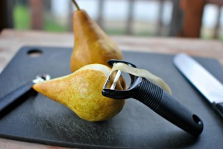 Pear peel extracts (Pyrus spp.)
“Total phenolic acids, total flavonoids, total triterpenes contents, and eleven monomeric compounds, derived from eight different pears’ peel and pulp, were qualitatively and quantitively profiled utilizing HPLC and HPLC-MS. All the chemical components found in the peel were approximately 2 to 18 folds concentrated than those in the pulp. Their antioxidant (reducing power and 2, 2-diphenyl-1-picrylhydrazyl/DPPH assay) and anti-inflammatory (xylene-induced mouse ear edema assay) activities were comparatively evaluated. Correlation analysis and principal component analysis were utilized to seek bioactive components in antioxidant and anti-inflammatory activities. Subsequently, Yaguang pear (Pyrus ussuriensis Maxim, with high polyphenols content) was selected to investigate in vitroand in vivo anti-diabetic potential (α-glucosidase inhibition assay and hypoglycemic assay in streptozocin-induced type 2 diabetic mice). Eleven monomeric compounds were seriatim examined for α-glucosidase inhibitory activity to fish out anti-diabetic components. For the first time, this study delineated pear peel’s anti-diabetic prospect of treatment for type 2 diabetes.”
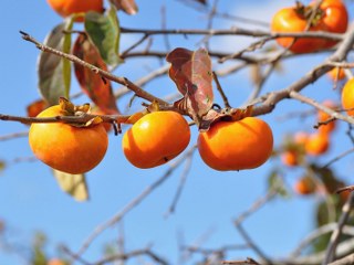 Persimmon peel extract
“The effect of persimmon peel polyphenol (PPP) on high glucose-induced oxidative stress was investigated using LLC-PK1 cells, which is susceptible to oxidative stress. High-concentration glucose (30 mM) treatment induced LLC-PK1 cell death, but high molecular-PPP (HMPPP) and low molecular-PPP (LMPPP), at concentrations of 5 or 10 μg/ml, significantly inhibited the high glucose-induced cytotoxicity. Furthermore, treatment with HMPPP or LMPPP dose-dependently reduced the intracellular reactive oxygen species level increased by 30 mM glucose. In addition, nitric oxide, superoxide and peroxynitrite levels were increased by 30 mM glucose treatment, but they were concentration-dependently inhibited by HMPPP or LMPPP treatment. High glucose levels induced the overexpressions of inducible nitric oxide synthase (iNOS) and cyclooxygenase-2 (COX-2) proteins, but HMPPP or LMPPP treatment reduced the overexpressions of these proteins. HMPPP or LMPPP also inhibited the nuclear translocation of nuclear factor-kappa B (NF-κB) induced by 30 mM glucose in LLC-PK1 cells. In particular, LMPPP exhibited stronger inhibitory activities on high glucose induced oxidative stress than HMPPP. These findings indicate the potential benefits of persimmon peel as a valuable source of antioxidants in the diabetic condition which will reduce the oxidative stress induced by hyperglycemia.”
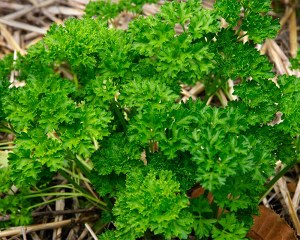 Petroselinum crispum extract
“Parsley is used by diabetics in Turkey to reduce blood glucose. The present study aims to investigate both the morphological and biochemical effects of parsley on liver tissue. Rat hepatocytes were examined by light and electron microscopy. Degenerative changes were observed in the hepatocytes of diabetic rats. These degenerative changes were significantly reduced or absent in the hepatocytes of diabetic rats treated with parsley. Blood glucose levels, alanine transaminase and alkaline phosphatase were observed to be raised in diabetic rats. Diabetic rats treated with parsley demonstrated significantly lower levels of blood glucose, alanine transaminase and alkaline phosphatase. The present study suggests that parsley demonstrates a significant hepatoprotective effect in diabetic rats.”
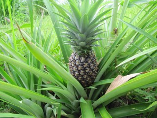
Pineapple (Ananas cosmosus) peel extract
“Oxidative stress was induced by oral administration of ethanol (20% w/v) at a dosage of 5 mL/kg body weight in rats. After 28 days of treatment, the rats were fasted overnight and sacrificed by cervical dislocation. Splenic tissue homogenates were used for the assessment of protein concentration, reduced glutathione (GSH), catalase, superoxide dismutase (SOD) and lipid peroxidation. Feeding on alcohol caused a significant decrease in GSH level in the group fed alcohol only, while treatment with pineapple peel extracts increased the GSH activities. No significant difference was observed in SOD levels of the negative control and group fed on only pineapple peel extract. There was an elevated catalase activity in the negative control. Pineapple peel extract significantly reduced it. Treatment with pineapple peel extracts was observed to reduce malondialdehyde (MDA) in alcohol‐fed groups. Protein concentration increased after treatment with the extracts. This study indicates the protective effect of pineapple peel extract against alcohol‐induced oxidative stress.”
 Pomegranate peel extract (Punica granatum )
“Peel extracts from Citrus sinensis, Punica granatum, and Musa paradisiaca were investigated for their effects on tissue lipid peroxidation (LPO) and on the concentration of thyroid hormones, insulin, and glucose in male rats. In vitro inhibition of H2O2-induced LPO in red blood cells of rats by 0.25, 0.50, 1.0, and 2.0 μg/mL C. sinensis, P. granatum, and M. paradisiacapeel extracts was observed in a dose-specific manner. Maximum inhibition was observed at 0.50 μg/mL C. sinensis, 2.0μg/mL P. granatum, and 1.0 μg/mL M. paradisiaca. In the in vivo investigation, out of four different concentrations of each peel extract, 25, 200, and 100 mg/kg C. sinensis, P. granatum, and M. paradisiaca, respectively, were found to maximally inhibit hepatic LPO. The most effective doses were further evaluated for effects on serum triiodothyronine (T3), thyroxine (T4), insulin, and glucose concentrations. C. sinensis exhibited antithyroidal, hypoglycemic, and insulin stimulatory activities, in addition to inhibition of LPO, as it significantly decreased the serum T4 (P < .05) and glucose (P < .001) concentrations with a concomitant increase in insulin levels (P < .05). P. granatum decreased LPO in hepatic, cardiac, and renal tissues (P < .01, P < .001, and P < .05, respectively) and serum glucose concentration (P < .01). M. paradisiaca strongly inhibited the serum level of thyroid hormones (P < .01 for both T3 and T4) but increased the level of glucose (P < .05). These findings reveal the hitherto unknown potential of the tested peel extracts in the regulation of thyroid function and glucose metabolism. Besides antiperoxidative activity, C. sinensis extract has antithyroidal, hypoglycemic, and insulin stimulatory properties, which suggest its potential to ameliorate both hyperthyroidism and diabetes mellitus.”
 Pomelo (citrus maxima) peel
“Metabolic syndrome is a serious health problem in both developed and developing countries. The present study investigated the anti-metabolic disorder effects of different pomelo varieties on obese C57BL/6 mice induced by high-fat (HF) diet.
The peels of four pomelo varieties were extracted with ethanol and the total phenols and flavonoids content of these extracts were measured. For the animal experiment, the female C57BL/6 mice were fed with a Chow diet or a HF diet alone or supplemented with 1% (w/w) different pomelo peel extracts for 8 weeks. Body weight and food intake were measured every other day. At the end of the treatment, the fasting blood glucose, glucose tolerance and insulin (INS) tolerance test, serum lipid profile and insulin levels, and liver lipid contents were analyzed. The gene expression analysis was performed with a quantitative real-time PCR assay.
The present study showed that the Citrus grandis liangpinyou (LP) and beibeiyou (BB) extracts were more potent in anti-metabolic disorder effects than the duanshiyou (DS) and wubuyou (WB) extracts. Both LP and BB extracts blocked the body weight gain, lowered fasting blood glucose, serum TC, liver lipid levels, and improved glucose tolerance and insulin resistance, and lowered serum insulin levels in HF diet-fed mice. Compared with the HF group, LP and BB peel extracts increased the mRNA expression of PPARα and its target genes, such as FAS, PGC-1α and PGC-1β, and GLUT4 in the liver and white adipocyte tissue (WAT).
We found that that pomelo peel extracts could prevent high-fat diet-induced metabolic disorders in C57BL/6 mice through the activation of the PPARα and GLUT4 signaling. Our results indicate that pomelo peels could be used as a dietary therapy and the potential source of drug for metabolic disorders.”
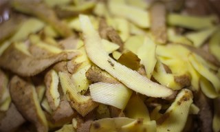 Potato Peel extract
“The potential of dietary potato peel (PP) powder in ameliorating oxidative stress (OS) and hyperglycemia was investigated in streptozotocin (STZ)-induced diabetic rats. In a 4-week feeding trial, incorporation of potato peel powder (5 and 10%) in the diet of diabetic rats was found to significantly reduce the plasma glucose level and also reduce drastically the polyuria of STZ diabetic rats. The total food intake was significantly reduced in the diabetic rats fed 10% PP powder compared to the control diabetic rats. However, the body weight gain over 28 days was nearly four times greater in PP powder supplemented diabetic rats (both at 5 and 10%) compared to the control diabetic rats. PP powder in the diet also decreased the elevated activities of serum transaminases (ALT and AST) and nearly normalized the hepatic MDA and GSH levels as well as the activities of specific antioxidant enzymes in liver of diabetic rats. The result of these studies clearly establishes the modulatory propensity of PP against diabetes induced alterations. Considering that potato peels are discarded as waste and not effectively utilized, these results suggest the possibility that PP waste could be effectively used as an ingredient in health and functional food to ameliorate certain disease states such as diabetes.”
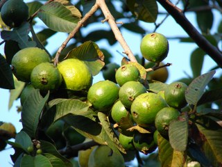 Psidium guajava raw fruit peel extract
Anti-hyperglycaemic potential of Psidium guajava raw fruit peel
“This study was undertaken to evaluate the glycaemic potential of aqueous extract of Psidium guajava unripe fruit peel on blood glucose level (BGL) of normal and streptozotocin induced mild and severely diabetic rats as an extension of our previous work carried out on Psidium guajava ripe fruit peel.
The aqueous extract of P. guajava unripe fruits was prepared. Male 6-8 wk old albino Wistar rats were selected for the experiments. Diabetes was induced by streptozotocin infection. Blood glucose levels were measured by glucose oxidase method. Antihyperglycaemic activity of the extract was assessed in mild and severely diabetic rats.
The maximum fall of 21.2 per cent (P<0.01) and 26.9 per cent (P<0.01) after 3 h of glucose administration during glucose tolerance test (GTT) was observed in BGL from a dose of 400 mg/kg, identified as the most effective dose, in normal and mild diabetic rats respectively. In severely diabetic rats the maximum fall of 20.8 and 17.5 per cent in fasting blood glucose (FBG) and post prandial glucose (PPG) levels, and 50 per cent (P<0.01) in urine sugar levels was observed with the same dose. Haemoglobin level increased by 5.2 per cent (P<0.05) and body weight by 2.5 per cent (P<0.05) after 21 days treatment.
Normal, mild and severely diabetic rat models had shown hypoglycaemic as well as antidiabetic effect of the unripe guava fruit peel aqueous extract. Further studies need to be done to characterize the active components of the peel.”
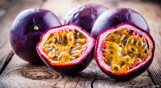 Purple passion fruit peel extract
The clinical efficacy of purple passion fruit peel extract (a flavonoid-rich dietary supplement) in reducing cardiovascular risk factors in adult type 2 diabetic subjects was investigated in a randomized, double-blind, placebo-controlled trial. Forty-one subjects were randomly assigned to receive a daily dose of purple passion fruit (220 mg) or a matched placebo for 16 weeks. Body mass index, blood pressure, fasting and postprandial blood glucose, glycated hemoglobin, and lipid profile were determined at baseline and at monthly intervals. A significant reduction in systolic blood pressure and fasting blood glucose was observed following administration of purple passion fruit (P<.05). Purple passion fruit was well tolerated, and no adverse events were reported. These data suggest that purple passion fruit supplementation for 16 weeks in type 2 diabetics results in a significant reduction in systolic blood pressure and fasting blood glucose, indicating that purple passion fruit is safe and well tolerated by diabetics.
 Quince (Cydonia oblonga Miller) peel extract
“Chronic inflammation is a hallmark of several pathologies, such as rheumatoid arthritis, gastritis, inflammatory bowel disease, atherosclerosis and cancer. A wide range of anti-inflammatory chemicals have been used to treat such diseases while presenting high toxicity and numerous side effects. Here, we report the anti-inflammatory effect of a non-toxic, cost-effective natural agent, polyphenolic extract from the Tunisian quince Cydonia oblongaMiller. Lipopolysaccharide (LPS) treatment of human THP-1-derived macrophages induced the secretion of high levels of the pro-inflammatory cytokine TNF-α and the chemokine IL-8, which was inhibited by quince peel polyphenolic extract in a dose-dependent manner. Concomitantly, quince polyphenols enhanced the level of the anti-inflammatory cytokine IL-10 secreted by LPS-treated macrophages. We further demonstrated that the unexpected increase in IL-6 secretion that occurred when quince polyphenols were associated with LPS treatment was partially responsible for the polyphenols-mediated inhibition of TNF-α secretion. Biochemical analysis showed that quince polyphenols extract inhibited the LPS-mediated activation of three major cellular pro-inflammatory effectors, nuclear factor-kappa B (NF-κB), p38MAPK and Akt. Overall, our data indicate that quince peel polyphenolic extract induces a potent anti-inflammatory effect that may prove useful for the treatment of inflammatory diseases and that a quince-rich regimen may help to prevent and improve the treatment of such diseases.”
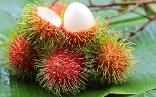 Rambutan (Nephelium lappaceum L.) Peel Extract
“Rambutan peel phenolic (RPP) extracts were prepared via dynamic separation with macroporous resin. The total phenolic content and individual phenolics in RPP were determined. Results showed that the total phenolic content of RPP was 877.11 mg gallic acidequivalents (GAE)/g extract. The content of geranin (122.18 mg/g extract) was the highest among those of the 39 identified phenolic compounds. RPP protected against oxidative stress in H2O2-induced HepG2 cells in a dose-response manner. The inhibitory effects of RPP on cell apoptosis might be related to its inhibitory effects on the generation of intracellular reactive oxygen species and increased effects on superoxide dismutase activity. The in vivo anti-aging activity of RPP was evaluated using an aging mice model that was induced by d-galactose (d-gal). The results showed that RPP enhanced the antioxidativestatus of experimental mice. Moreover, histological analysis indicated that RPP effectively reduced d-gal-induced liver and kidney tissue damage in a dose-dependent manner. Therefore, RPP can be used as a natural antioxidant and anti-aging agent in thepharmaceutical and food industries.”
 Satsuma Mandarin peel extract (CitruS unshiu)
Lipolysis induced by segment wall extract from Satsuma mandarin orange
“The lipolysis induced by Satsuma mandarin orange (Citrus unshu Mark) was investigated using rat fat cells. Peel or segment wall extract from Satsuma mandarin orange induced the lipolysis in a concentration-dependent manner, whereas juice sac extract did not induce the lipolysis. High concentration of synephrine, which is an adrenergic amine, was detected in the peel or segment wall extract, whereas it was not detected in the juice sac extract. The segment wall extracts from Iyokan and orange had high lipolytic activity, whereas the extracts from grapefruit and lemon did not have lipolytic activity. The beta-antagonist inhibited the lipolysis elicited by the segment wall extract from Satsuma mandarin orange, whereas alpha-antagonist did not inhibit the lipolysis induced by the segment wall. The lipolysis induced by the segment wall was considerably higher in the visceral fat cells when compared to the subcutaneous fat cells. These results suggest that the segment wall, an edible fraction, from Satsuma mandarin orange might be useful as a functional food, especially as a fat-reducing material.”
 Scutellaria laterifolia
“Traditional Chinese medicines have been recently recognized as a new source of anticancer drugs and new chemotherapy adjuvant to enhance the efficacy of chemotherapy and to ameliorate the side effects of cancer chemotherapies however their healing mechanisms are still largely unknown. Scutellariabaicalensis is one of the most popular and multi-purpose herb used in China traditionally for treatment of inflammation, hypertension, cardiovascular diseases, and bacterial and viral infections. Accumulating evidence demonstrate that Scutellariaalso possesses potent anticancer activities. The bioactive components of Scutellaria have been confirmed to be flavones. The major constituents of Scutellaria baicalensis are Wogonin, Baicalein and Baicalin. These phytochemicals are not only cytostatic but also cytotoxic to various human tumor cell lines in vitro and inhibit tumor growth in vivo. Most importantly, they show almost no or minor toxicity to normal epithelial and normal peripheral blood and myeloid cells. The antitumor functions of these flavones are largely due to their abilities to scavenge oxidative radicals, to attenuate NF-κB activity, to inhibit several genes important for regulation of the cell cycle, to suppress COX-2 gene expression and to prevent viral infections. The tumor-selectivity of Wogonin has recently been demonstrated to be due to its ability to differentially modulate the oxidation–reduction status of malignant vs. normal lymphocytic cells and to preferentially induce phospholipase Cγ1, a key enzyme involved in Ca2+ signaling, through H2O2 signaling in malignant lymphocytes. This review is aimed to summarize the research results obtained since the last 20 years and to highlight the recently discovered molecular mechanisms.”
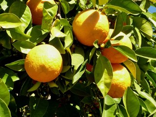
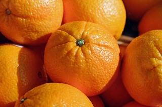
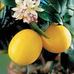
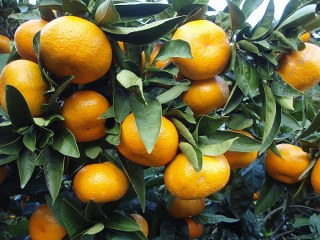
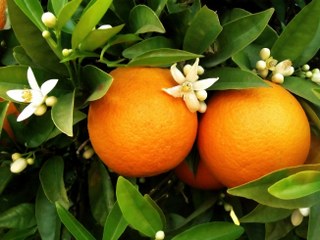 Sweet Orange peel extract (Citrus sinensis)
“Orange (citrus sinensis) is well known for its nutritional and medicinal properties throughout the world. From times immemorial, whole Orange plant including ripe and unripe fruits, juice, orange peels, leaves and flowers are used as a traditional medicine. Citrus sinensis belongs to the family Rutaceae. The fruit is a fleshy, indehiscent, berry that ranges widely in size from 4 cm to 12 cm. The major medicinal properties of orange include anti-bacterial, anti-fungal, anti- diabetic, cardio- protective, anti-cancer, anti-arthritic, anti-inflammatory, antioxidant, anti-Tubercular, anti-asthmatic and anti-hypertensive. Phytochemically, whole plant contains limonene, citral, neohesperidin, naringin, rutin, rhamnose, eriocitrin, and vitamin-C. In the present review article, a humble attempt is made to compile all the strange facts available About this tasty fruit.
Anti-diabetic activity of orange is due to bioflavonoids such as hesperidin and naringin present in citrus fruit peels. These peels play an anti-diabetic rolein C57BL/Ks J-db/db mice via regulation of glucose regulatory enzymes. They decrease the activity of glucose-6-phosphatase and phosphoenol pyruvate. The anti-diabetic potential of orange peel and juice appear to be mediated via anti peroxidation, inhibition of α-amylase enzyme activity that is responsible for the conversion of complex carbohydrates to glucose, increased hepatic glycogen content, stimulation of insulin secretion, and repair of secretory defects of pancreatic β-cells [18,19].”
 Syzygium cumini Skeels extract
Syzygium cumini (L.) Skeels: A review of its phytochemical constituents and traditional uses
“Syzygium cumini (S. cumini) (L.) Skeels (jambolan) is one of the widely used medicinal plants in the treatment of various diseases in particular diabetes. The present review has been primed to describe the existing data on the information on botany, phytochemical constituents, traditional uses and pharmacological actions of S. cumini (L.) Skeels (jambolan). Electronic database search was conducted with the search terms of Eugenia jambolana, S. cumini, jambolan, common plum and java plum. The plant has been viewed as an antidiabetic plant since it became commercially available several decades ago. During last four decades, numerous folk medicine and scientific reports on the antidiabetic effects of this plant have been cited in the literature. The plant is rich in compounds containing anthocyanins, glucoside, ellagic acid, isoquercetin, kaemferol and myrecetin. The seeds are claimed to contain alkaloid, jambosine, and glycoside jambolin or antimellin, which halts the diastatic conversion of starch into sugar. The vast number of literatures found in the database revealed that the extracts of different parts of jambolan showed significant pharmacological actions. We suggest that there is a need for further investigation to isolate active principles which confer the pharmacological action. Hence identification of such active compounds is useful for producing safer drugs in the treatment of various ailments including diabetes.”
Tangerine peel extract (Citrus reticulata)
“Naringin and naringenin supplementation has proven to be efficacious for the treatment of metabolic syndrome and obesity in animal models (60, 111). The dose used in animal studies may not be achieved in human trials, however, and emphasis should be given to establish a dietary recommendation of citrus fruit consumption or for the pure compound naringin. Most of naringin’s observed biologic activities are related in some part to its potent antioxidant nature. However, several other molecular signaling pathways modified by naringin were also revealed recently. In metabolic syndrome, obesity, and related cardiovascular complications, naringin influences AMPK-, PPARα–, and CPT-1–mediated fat utilization and preserves mitochondrial function. Moreover, naringin also prevents the TNF-α–mediated inflammatory process and tissue damage in liver and vasculature. Figure 3 shows a proposed mechanism for increased mitochondrial biogenesis by naringin. Another consideration for oral administration of naringenin/naringin should be the contraindications of taking certain drugs such as statins and calcium channel blockers because of the inhibitory effects on intestinal CYP450 enzymes. Moreover, further clinical investigations should be carried out with naringin in metabolic diseases.”
 Tomato (Solanum lycopersicum L.) peel extract
“Lycopene rich extracts (ETE) were prepared by enzyme assisted extraction of tomato peel. Oxidative damage was induced in L6 myoblasts by exposing cells to H2O2 in presence and absence (control) of ETE. The potential of ETE to protect cells from oxidative damage was investigated in terms of cytotoxicity (MTT assay), Reactive oxygen species (ROS) by 2′, 7′-dichlorofluorescindiacetate (H2DCFDA), DNA damage (ladder assay, 8-oxo-dG), Hoechst 33342 nuclear staining, mitochondrial stability by ATP production and mitochondrial membrane potential by Rhodamine 123 staining. It was observed that treatment of cells with ETE significantly increased cell viability, decreased ROS production, reduced DNA fragmentation, chromatin condensation & 8-oxo-dG, increased ATP levels and mitochondrial membrane potential. Results indicated that ETE could protect cells from H2O2 induced oxidative damage significantly as compared to control. These results depicted the antioxidant potential of tomato peel extract which can counteract the redox imbalance in cells induced by oxidative stress condition.”
 Trapa natans fruit peel extract
“Trapa natans L. (Lythraceae), commonly known as Water Chestnut in English, is an annual aquatic floating herb occurring throughout the Indian subcontinent and used traditionally for several medicinal purposes. The present study was aimed to evaluate antidiabetic activity of methanol extract of T. napans fruit peels (METN) in Wistar rats. The effect of METN on oral glucose tolerance and its effect on normoglycemic rats were studied. Diabetes mellitus was induced in rats by single intraperitonial injection of streptozotocin (STZ, 65 mg/kg body weight). Three days after STZ induction, the hyperglycemic rats were treated with METN orally at the dose of 100 and 200 mg/kg body weight daily for 15 days. Glibenclamide (0.5 mg/kg b.wt., orally) was used as reference drug. The fasting blood glucose levels were measured on every 5 day during the th 15 days treatment. METN at the dose of 100 and 200 mg/kg orally significantly (p < 0.001) and dose dependently improved oral glucose tolerance, exhibited hypoglycaemic effect in normal rats and antidiabetic activity in STZ-induced diabetic rats by reducing and normalizing the elevated fasting blood glucose levels as compared to those of STZ control group. The present study concludes that T. natans fruit peel demonstrated promising antidiabetic activity in STZ-induced diabetic Wistar rats.”
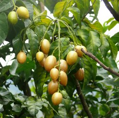 Wampee (Clausena lansium (Lour.) Skeels) Peel extract
“Fruits of wampee [Clausena lansium (Lour.) Skeels] contain a significant amount of coumarins with many health benefits. The activity-guided separation of an ethyl acetate-soluble fraction on a polyamide column followed by silica gel column and high performance liquid chromatography (HPLC) preparation afforded a pure compound, which was identified to be 8-hydroxypsoralen based on the 1H, 13C NMR (nuclear magnetic resonance), and ESI-MS (electrospray ionisation mass spectrometric) analysis. This isolate exhibited good scavenging activities against DPPH radical and superoxide anion as well as significant reducing power. It also showed potent proliferation inhibitory activity against human hepatocellular liver carcinoma cell line (HepG2), human lung adenocarcinoma epithelial cell line (A549) and human cervical carcinoma cell line (HELA). This is the first report on the antioxidant and cytotoxic properties of C. lansium fruit extract. The food and pharmaceutical industry could be benefited by the usage of this extract containing this constituent.”
Water chestnut (Trapa bispinosa Roxb.) peel
“In recent years, glycation in organisms has been positioned as a major risk factor for accelerated aging process, and the concept of glycation stress has gained attention 1). The concept of glycation stress is that the reaction of reducing sugars, aldehydes, etc. with proteins and amino acids produces advanced glycation end products (AGEs), and the accumulation of these substances overloads cells and tissues and disrupts the functioning of functional proteins as well as causes a series of reactions such as induction of inflammation 2). The glycation reaction at the core of glycation stress is a reaction in which proteins and reducing sugars bond nonenzymatically to produce glycation products. Glycation products that have been identified include the AGEs pentosidine, Nε- (carboxymethyl ) lysine (CML), and pyraline, and their intermediates 3-deoxyglucosone (3DG), glyoxal (GO), and methylglyoxal (MGO) 3). Bioaccumulation of these glycation products is said to be involved in kidney disease and eye disorders associated with progression of diabetes 4), arteriosclerosis 5), osteoporosis 6), Alzheimer’s disease 7), infertility 8), and sclerema 9). Based on these facts, we believe that inhibiting the production and accumulation of glycation products and stimulating the degradation and metabolism of previously produced glycation products will lead to an increased life expectancy, improved quality of life, and disease prevention.
The glycation response inhibiting activity and glycation product degradation activity of the TBE confirm its production inhibitory effect against AGEs and its effect in reducing the amount of accumulated AGEs by degradation of previously produced AGEs. Further, since activities other than antiglycation have been demonstrated, we expect TBE to have wide applications as an anti-aging ingredient. We plan to study its efficacy in humans in the future. The study verified TBE to inhibit glycation product synthesis, degrade α-dicarbonyl compound, and break the AGE crosslinks. The above results suggest that TBE may inhibit production of AGEs and their intermediates and degrade existing α-dicarbonyl compounds and AGE crosslinks in vivo. We expect that TBE may be suitable to reduce glycation stress.”
 Yam (Dioscorea alata) peel extract
“Researchers have shown that yam extracts contain antioxidative activity; however, there are few reports regarding the antioxidant activities of yam peel. The effects of water and 50% ethanolic extracts from Darsan yam (Dioscorea alata) peel on the oxidative status of tert-butylhydroperoxide (t-BHP)-treated mouse Hepa 1–6 and FL83B liver cell lines were investigated. The cytosols were analysed for H2O2 and malondialdehyde (MDA) levels and antioxidative enzymes activities, including superoxide dismutase, glutathione peroxidase (GPx) and catalase activities. Both water and 50% ethanolic extracts from yam peel did not affect cellular MDA level in t-BHP-treated cells, but they altered the level of H2O2. Water extract from yam peel amplified the t-BHP-induced cytotoxicity in Hepa 1–6 whilst the ethanolic extract showed protection in FL83B cells. GPx activity might play an important role in the protective effect associated with t-BHP-induced oxidative stress.”

Yuzu peel extract (Citrus JUNOS)
“The antidiabetic effect of the Citrus junos Tanaka (also known as yuja or yuzu) was examined. Ethanol extract of yuja peel (YPEE) significantly stimulated 2-[N-(7-nitrobenz-2-oxa-1,3-diazol-4-yl)amino]-2-deoxy-D-glucose (2-NBDG) uptake in C2C12 myotubes. However, ethanol extract of yuja pulp (YpEE) and water extract of yuja peel (YPWE) or pulp (YpWE) did not stimulate glucose uptake. In addition, peroxisome proliferator-activated receptor gamma (PPAR-) and AMP-activated protein kinase (AMPK) activities were increased by YPEE in a dose-dependent manner. Pretreatment of AMPK inhibitor decreased the glucose uptake stimulated by YPEE in C2C12 myotubes. We confirmed the anti-diabetic effect of YPEE in mice fed a high fat-diet (HFD). Compared with control mice on a normal diet (ND), these mice showed increased body weight, liver fat, insulin resistance, triacylglycerol (TG), and total cholesterol content. Addition of 5% YPEE significantly reduced the weight gain and rise in liver fat content, serum triacylglycerol (TG), total cholesterol, and insulin resistance found in mice fed a high-fat diet (HFD). Moreover, YPEE reduced the secretion of HFD-induced adipocytokines such as leptin and resistin. YPEE also resulted in increased phosphorylation of AMPK in muscle tissues. These results suggest that ethanol extract of yuja peel exerts anti-diabetic effects via AMPK and PPAR- in both cell culture and mouse models.”
AVAILABLE IN TWO CONCENTRATIONS: 20:1 100g $50 / 200:1 100g $250
| Concentration | 20:1, 200:1 |
|---|
Related products
-
LUTEOLIN 200:1
$442.00
Reviews
There are no reviews yet.